What the GI-MAP Test Actually Shows — And Why It Finds What Dubai Doctors Miss
The GI-MAP stool test measures what standard UAE gastroenterology panels skip. Here is what it actually detects and why it matters.
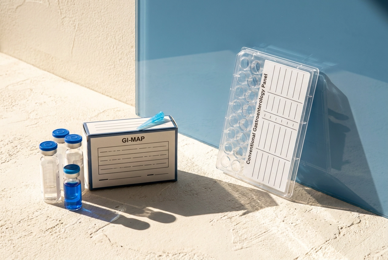
You have had the scopes, the bloods, the ultrasounds. Every result has come back unremarkable, and yet your digestion is unreliable, your energy is inconsistent, and something clearly is not right. This is one of the most common clinical patterns seen in women seeking functional medicine support across the UAE, and it has a straightforward explanation: the conventional gastroenterology workup was not designed to find what is actually driving your symptoms.
The GI-MAP test, available to clients in Dubai and across the Emirates through specialist functional medicine practitioners, operates on an entirely different diagnostic layer. Using quantitative PCR technology, it identifies and quantifies pathogens, opportunistic bacteria, commensal populations, parasites, H. pylori virulence factors, intestinal inflammation markers and immune function indicators from a single stool sample. Standard panels simply do not go here.
This analysis walks through exactly what conventional GI testing measures and where its blind spots are, how the GI-MAP works, what each marker category reveals, and why the newest StoolOMX add-on is changing what is clinically actionable for women who have run out of conventional answers.

Normal results, still feel terrible
The report is in your hand. It says no pathology found. Your abdomen is visibly distended and you have not had a complete bowel movement in four days.
That is not a mystery. That is a measurement gap.
As covered below, conventional panels ask only whether an acute pathogen is present; the GI-MAP asks what the gut environment actually looks like. Applied to a woman with chronic, low-grade, multi-system digestive dysfunction, a standard panel is simply the wrong instrument. Not because the gastroenterologist missed something. Because the test was never designed to see it.
Standard panels ask only whether an acute pathogen is present (explored in the next section); the GI-MAP asks what the gut environment actually looks like: the microbial balance, the immune layer, the digestive capacity, the inflammatory activity. That is terrain assessment, not pathology detection. Three things most doctors never test for illustrates exactly how far outside standard panels these markers sit.
The goal is not a new diagnosis to match to a new pill. It is the data layer that tells you what the terrain requires before you build a protocol. That distinction matters more than any single result on the report.
What conventional panels actually test, and what they skip

Standard hospital stool cultures are built for one purpose: ruling out acute infection. The panel typically targets a short list of common bacterial pathogens and parasites. If none of those are present, the report reads negative, and negative gets communicated as normal.
The methodology compounds this. These tests are qualitative. Every organism either clears a detection threshold or it does not. There is no number attached. You cannot tell whether a commensal species has overgrown into an opportunistic load, because the test was never designed to quantify. A culture that returns negative on Klebsiella tells you nothing about whether Klebsiella is present at a clinically irrelevant trace level or at a high colonisation load disrupting the entire ecosystem around it.
What the panel omits is the more significant gap. Intestinal inflammation markers, mucosal immune status, digestive enzyme output, gut barrier integrity: none of these typically appear on a standard gastroenterology stool culture. None of them require an acute infection to be compromised. They can be significantly disrupted in a woman who has never had food poisoning in her life.
H. pylori is worth naming specifically. A standard stool culture confirms H. pylori presence or absence. It does not report virulence gene status. That distinction changes the entire picture, and it is invisible on conventional testing.
The result is a panel designed for emergency and infectious disease medicine, applied to a clinical presentation it was never built to assess.
How the GI-MAP works
The GI-MAP (Gastrointestinal Microbial Assay Plus) uses quantitative PCR on a single stool sample. One collection, one shipment, results in 7 to 10 business days.
The quantitative part is what separates it from the panels described above. Standard cultures return a positive or a negative. PCR measures how much of each organism's DNA is present, expressed as a numerical load. That distinction is not a technical footnote; it changes clinical meaning entirely. Candida at trace levels in an otherwise intact microbiome is not the same clinical picture as Candida at high colonisation in a depleted one. The number determines the relevance.
PCR also detects organisms that do not survive the journey through standard culture conditions. Many parasites, anaerobic bacteria, and slow-growing fungi require live, viable cells to show up on a culture plate. If the organism dies in transit or simply does not grow on standard media, the culture returns negative regardless of what is actually present. PCR reads DNA, not viability.
One validation point worth naming honestly: a peer-reviewed study comparing the GI-MAP to the BioFire FilmArray reference panel found 80% sensitivity but only 26% specificity for pathogen detection, meaning a meaningful rate of false positives in that specific comparison. That is a real limitation for its pathogen arm. It is also a narrow frame. The BioFire panel does not measure mucosal immune status, intestinal inflammation, digestive enzyme output, commensal populations, or H. pylori virulence genes. The GI-MAP's clinical weight sits precisely in those categories, which the reference panel was never designed to assess and cannot speak to at all.
That is where the next section picks up.
The marker categories that change the picture
Those markers the reference panel does not measure, that is where the clinical picture lives.
Pathogens covers the expected ground: bacterial targets including Clostridioides difficile, enterotoxigenic E. coli, and Campylobacter; parasites including Giardia, Cryptosporidium, and Entamoeba histolytica; viral targets including Adenovirus 40/41 and Norovirus; and fungal organisms. One panel, one sample.
Opportunistic bacteria is where it gets more interesting. Klebsiella pneumoniae, Pseudomonas aeruginosa, Staphylococcus aureus, these organisms exist in many healthy guts at low, unremarkable loads. Quantity changes everything. When the commensals that normally keep them in check are depleted, their loads climb, and they shift from bystanders to contributors to inflammation and dysbiosis. A qualitative panel cannot see this because it does not measure load.
Commensal bacteria are not tested at all on standard panels. The GI-MAP quantifies Lactobacillus, Bifidobacterium, Akkermansia muciniphila, and Faecalibacterium prausnitzii. These species determine how resilient the terrain is. Low Faecalibacterium prausnitzii is a marker of reduced ecosystem resilience. You cannot restore what you have not measured.
H. pylori virulence factors change the clinical significance of a positive result entirely. The GI-MAP reports virulence genes: CagA, VacA, DupA, IceA, OipA. A CagA-positive strain carries a meaningfully higher risk profile than a CagA-negative one. "H. pylori detected" without virulence typing is incomplete information.
Intestinal inflammation markers: calprotectin is a marker of active mucosal inflammation. Anti-gliadin IgA measures an immune response to gliadin.
Secretory IgA (SIgA) is the primary mucosal antibody. Chronically low SIgA means impaired pathogen clearance and poor immune tolerance. Chronically elevated SIgA signals ongoing immune activation. Both patterns are clinically significant. Neither appears on a standard panel.
Elastase-1 reflects pancreatic exocrine output. When it is low, the pancreas is not producing adequate digestive enzymes. Steatocrit reflects fat malabsorption alongside it.
Beta-glucuronidase is a bacterial enzyme involved in estrogen metabolism in the gut. For any woman presenting with hormonal symptoms alongside digestive ones, this marker is directly relevant. It does not appear on a standard stool culture.
The StoolOMX layer: bile acids and short-chain fatty acids
All of that is happening at the microbial and immune level. The StoolOMX add-on goes one layer deeper: it measures the metabolic output of those microbial populations.
The panel profiles bile acids and a range of short-chain and branched-chain fatty acids (SCFAs), giving a functional readout that conventional stool testing does not include.
SCFAs, primarily butyrate, propionate, and acetate, are produced when gut bacteria ferment fibre. Butyrate is the primary fuel source for colonocytes and a key regulator of intestinal barrier integrity. When butyrate-producing species are depleted, the epithelial layer is not being fed. That is a mechanism driving barrier compromise, not a symptom of it. You cannot see it on an endoscopy or infer it from bloating patterns.
Bile acid profiling adds a second dimension. Primary bile acids are synthesised in the liver; gut bacteria transform them into secondary bile acids in the colon through deconjugation and other chemical modifications. The bile acid profile reflects which microbial populations are performing that transformation and connects the metabolite picture back to the microbiome data.
Research on microbiota-SCFA relationships confirms that SCFA composition tracks with clinical heterogeneity in ways symptom reporting cannot distinguish. The metabolite layer is what separates knowing the terrain is dysfunctional from knowing what it is actually doing.
Why this matters specifically for women getting GI workups in the UAE
None of that data gap is your doctor's fault. A gastroenterologist in Dubai is running the tests the system provides. Those tests were built to answer one question: is there an acute, reportable pathogen? When the answer is no, the panel has done exactly what it was designed to do. It cannot report on markers it does not include.
That structural mismatch hits women harder. The markers that most frequently explain female-pattern presentations, low SIgA, elevated beta-glucuronidase, suppressed elastase-1, depleted commensals, are not part of a standard stool culture or endoscopy workup. They are not missed through oversight. They are simply outside the scope of what the test was built to find.
For women in a pre-conception window, the stakes compound. Accessing a GI-MAP test does not require a hospital referral, functional medicine practitioners can order the test and work with clients on results remotely, with collection completed from a single home stool sample.
What it returns is not a diagnosis. It is a terrain map. That distinction matters because a map sequences a protocol correctly rather than sending you back through another round of restriction or targeted killing that addresses a branch while the root stays intact.
What the data enables: restoration, not restriction
That data map is only useful if you know what to do with it. Most practitioners use it as a treatment target list: high pathogen load, add an antimicrobial; low Lactobacillus, add a probiotic. That sequence feels logical. It is also why symptoms come back.
The GI-MAP does not tell you what to eliminate. It tells you what the terrain requires before any intervention will hold.
As the marker section established, depleted keystones signal terrain unreadiness, not a simple probiotic gap. When elastase-1 is low, partially digested food reaches the colon and feeds the same dysbiotic populations you are trying to clear, running a full antimicrobial protocol may shift the microbial picture temporarily, but without addressing that feeding condition, dysbiosis rebounds. When SIgA is low, the mucosal immune layer is not in a position to support new colonisation.
This is why, at Satori, the GI-MAP runs alongside three other labs at the start of every personalised protocol, not as a standalone test: the OAT for organic acids and mitochondrial function, the DUTCH for hormone and cortisol metabolites, and the HTMA for mineral status and heavy metal burden. Together they produce a Root Cause Map that sequences what the body needs and in what order.
That order is not arbitrary. Drainage, stomach acid and bile, motility, minerals, nervous system regulation, and gut lining integrity all come before any antimicrobial phase. Not because killing is wrong. Because killing into an unprepared terrain is precisely why the last protocol did not hold.
If your results have been normal and you still feel this way
You now have the vocabulary for what was missing. Commensal depletion, suppressed mucosal immunity, impaired pancreatic output, disrupted bile acid metabolism, low butyrate production, these are measurable, specific, and correctable. They do not require a diagnosis. They require data.
Calprotectin, bile acids, SCFAs, none of these appeared on your panel because none were tested. A clean hospital report and a symptomatic gut are not contradictory findings. They are what happens when the wrong question gets asked with a well-calibrated instrument.
If you want to know which of these systems is most likely driving your pattern, the free Root Cause Assessment at myrootcause.satorihealth.co is the starting point. It maps your symptom picture to the marker categories most likely to explain it. No protocol, no pressure. Just a clearer picture of where to look first.
You are not broken. You are under-measured. That is a solvable problem.
Conclusion
Your symptoms are not imaginary, and your normal results are not the final word. Conventional GI panels test for acute pathogens and leave the deeper architecture of gut function completely unmeasured. The GI-MAP, paired with the StoolOMX layer, fills that gap by quantifying commensals, mucosal immunity, digestive output, bile acid metabolism, and short-chain fatty acid production.
This is the difference between asking whether something is wrong and asking how the system is actually functioning.
As established throughout, if you have been handed a clean report that explains nothing, the free Root Cause Assessment at myrootcause.satorihealth.co is the logical next step. It takes your symptom picture and maps it to the marker categories most likely driving it.